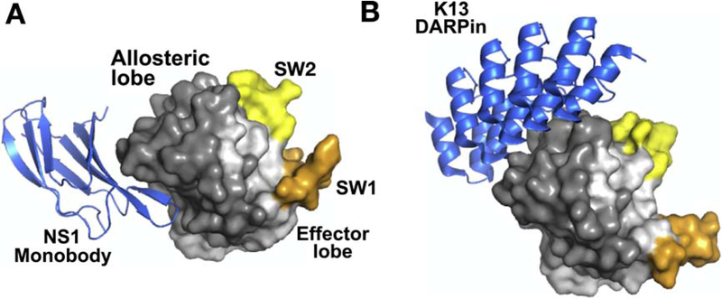
Figure 5. Biologics targeting RAS.
A. Structure of NS1 monobody bound to HRAS WT (PDB:5E95). B. Structure of DARPin K13 bound to KRAS(G12V):GDP (PDB: 6H46). Both structures are shown in the same orientation to illustrate the different regions targeted by NS1 versus K13. Coloring of figure is the same as in Fig. 4. Both NS1 and K13 target the allosteric lobe of RAS.

