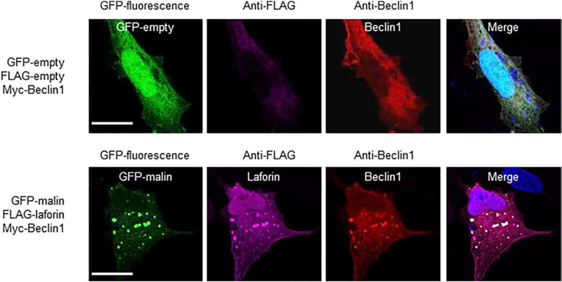
Fig. 3.-. Laforin and malin co-localize with Beclin1.
Representative images of U2OS cells transfected with the plasmids indicated on their left. The subcellular localization of the corresponding proteins was analyzed either by direct fluorescence (GFP and GFP-malin) or by immunofluorescence using anti-FLAG (coupled to an Alexa-Fluor 633 secondary antibody) and anti-Beclin1 (coupled to an Alexa-Fluor 568 secondary antibody) antibodies, as described in Materials and Methods. Fluorescence associated with the corresponding proteins was analyzed by confocal microscopy. DAPI staining was in blue. A merged image of the fluorescent dyes is also shown. Bar: 20 μm.

