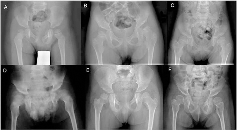
Figure 3:

Pelvic radiographs in late infantile patients show hypoplasia of the infero-lateral portions of the ilia resulting in slanted acetabular roofs and wide ileo-acetabular angles. A. Patient GSL022, 3 years. The femoral necks are in valgus. The capital femoral epiphyses are well rounded and slightly large. B. Patient GSL023, 5 years. C. Patient GSL007, 6 years. D. Patient GSL072, 8 years. E. Patient GSL013, 4 years. The capital femoral epiphyses are dysplastic due to deficient ossification of their medial portions. F. Patient GSL013, 9 years. Compared to E, epiphyseal ossification has normalized, and the femoral necks are shorter.
