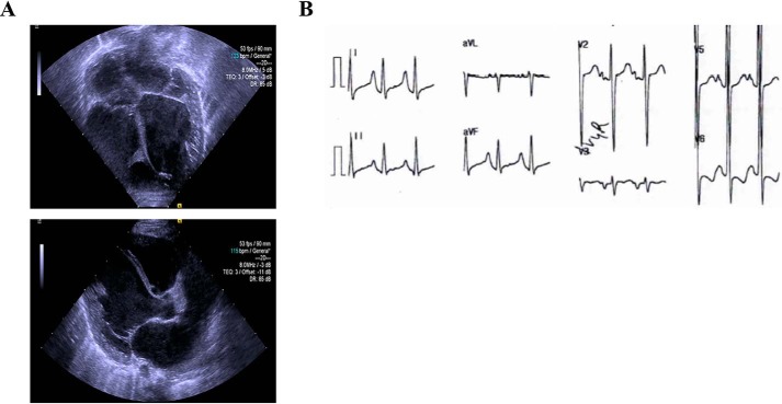
Figure 1.
Clinical presentation of the proband. A, representative echocardiographic images demonstrating left ventricular dilation and hypertrabeculation (top); parasternal long axis view showing dilation of the left atrium and left ventricle (bottom). B, representative electrocardiogram tracings showing sinus rhythm with criteria satisfied for biventricular hypertrophy, right atrial enlargement, and diffuse STT changes.

