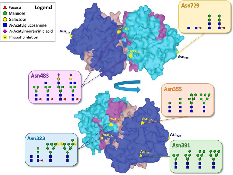
Figure 1.
Summary of the major glycan compositions found on each N-glycosylation site of healthy human myeloperoxidase (MPO). The light and dark blue regions represent the two heavy chains within the MPO homodimer, the brown and purple regions the light chains. The structure corresponds to the PDB entry 1CXP (32).

