Figure 3.
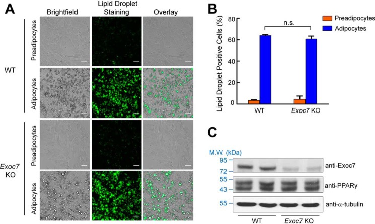
Inducible KO of Exoc7 permits normal adipocyte differentiation. A, representative images of preadipocytes and adipocytes. Dox was added to adipocytes but not preadipocytes. Lipid droplets in the cells were stained with Nile red, and the images were captured using a ×20 objective on an Olympus IX81 microscope. Scale bars = 50 μm. B, quantification of lipid droplet–positive cells (WT or Exoc7 KO). Cells were stained with Nile red and the fluorescence of Nile red was measured using flow cytometry. Data are presented as mean ± S.D. n = 3. n.s., not significant, p > 0.05. C, representative immunoblots showing expression of the indicated proteins in WT and Exoc7 KO adipocytes. Two independent samples were prepared for each cell line. M.W., molecular weight.

