Figure 5.
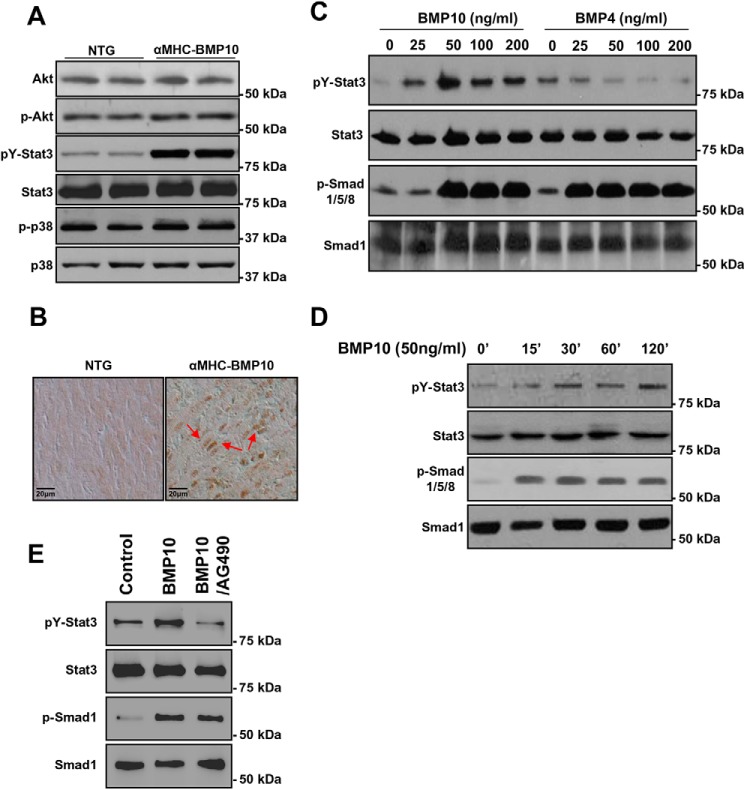
Biochemical analysis of BMP10-mediated activation of STAT3. A, Western blotting analysis of key signaling molecules in TG and NTG hearts showing STAT3 was significantly activated. B, immunohistochemical analysis of the subcellular localization of STAT3 in TG and NTG hearts, with positive nuclear STAT3 staining (red arrows) indicating the activation of STAT3 and its mediated signaling pathway. C, Western blotting analysis of STAT3 activation in P19 cells in response to rhBMP10 and rhBMP4 at different concentrations compared with Smad activation. D, Western blotting analysis of the time course of STAT3 activation by BMP10. E, Western blotting analysis of STAT3 activation in response to BMP10 and JAK1 inhibitor AG490. After overnight starvation, the cells are treated with rhBMP10 (50 ng/ml) with either DMSO or AG490 (5 μm in DMSO). The quantification of the Western blotting is shown in Fig. S3.

