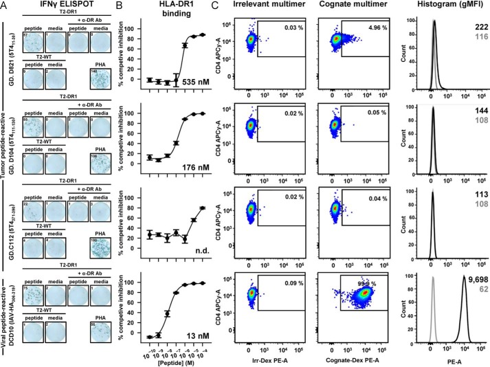
Figure 2.
5T4 clone reactivity to HLA-DR1–presented peptides despite no measurable ligand engagement. A, IFN-γ ELISpot assays of clones in response to overnight co-incubation with peptide-pulsed APCs. IFN-γ release was observed using DR1-only (T2-DR1) but not DR1-null (T2-WT) presenters in the presence of peptide but not no-peptide (media) controls. IFN-γ release was blocked by an αDR blocking antibody (+α-DR Ab). GD.D821, GD.D104, GD.C112, and DCD10 were stimulated with APCs pulsed with 10−5, 10−6, 10−5, and 10−6 m peptide, respectively. Maximal IFN-γ response indicated by phytohemagglutinin (PHA) activation. Inset numbers represent raw spot forming cells (sfcs). Presented ELISpot wells representative of two duplicate experiments. B, binding capacity of each peptide to HLA-DR1 molecules in competitive binding assays in vitro. Error bars indicate S.D. of experiments performed in triplicate. Inset number denotes IC50 value calculated from displayed curve fit. N.D. = IC50 not determined. C, cognate HLA-DR1 multimer staining of each 5T4-reactive clone exhibiting staining marginally above background (irrelevant multimer). This was in stark contrast to typical staining of the DCD10 viral-reactive clone. Histogram representation displays inset geometric mean fluorescent intensity of cognate -DR1 multimer (black) and irrelevant -DR1 control (gray).

