Figure 4.
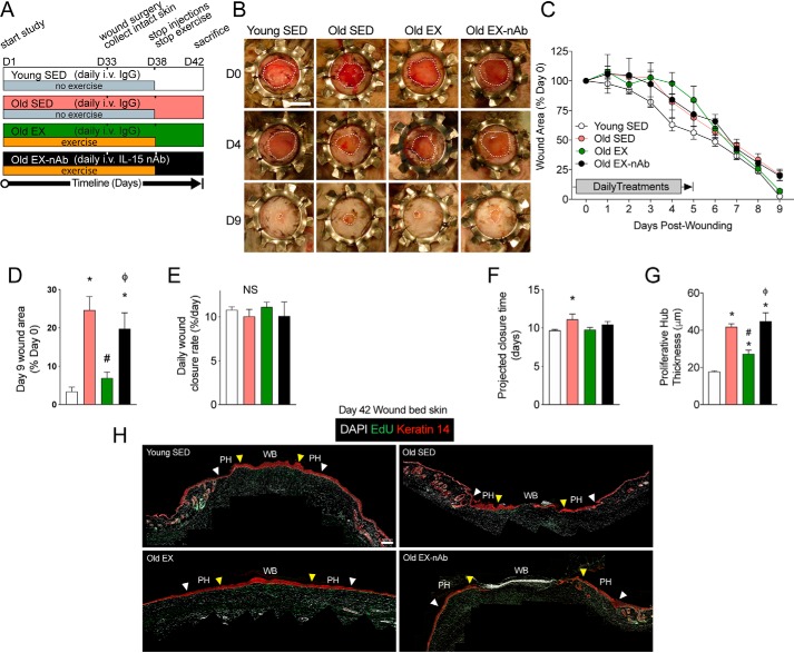
Exercise-induced improvements in wound healing depend on IL-15 signaling in aged mice. A, experimental design of intravenous pre-treatments, the exercise regimen, and wound surgery with key experimental days denoted on a timeline. B, representative images of wound healing progress in young sedentary (Young SED), old sedentary (Old SED), old exercised (Old EX), and old exercised mice receiving IL-15 neutralizing antibody (Old EX-nAb) on the indicated days post-wounding. Wound margins are delineated by the dotted white line. Scale bar = 5 mm. C, quantification of wound areas from B. D, final wound closure at time of sacrifice on day 9 expressed as a proportion of the original wound area. E, wound closure rate per day determined by average slope of best fit lines. F, projected time for full wound closure based on wound closure rate calculated in E. n = 4–8 mice per group in A–E. G, quantification of the proliferative hub thickness in each treatment group. n = 3–5 mice per group. H, stitched panoramic immunofluorescence images of Keratin 14-stained stem cells and EdU labeling in day 9 post-wounded skin showing the wound bed (WB) and proliferative hub (PH) region of each side. The space between the white arrow (original wound edge) and the yellow arrow (300 μm inwards) indicates the proliferative hub region. Scale bar = 100 μm. Data are mean ± S.E. *, significantly different (p < 0.05) relative to Young SED mice. #, significantly different (p < 0.05) relative to Old SED mice. ϕ, Significantly different (p < 0.05) relative to Old EX mice.

