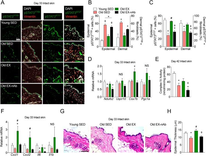
Figure 5.
Neutralization of IL-15 in exercising mice prevents the rescue of STAT3 phospho-signaling, mitochondrial and inflammatory signaling, and epidermal structure. A, representative immunofluorescence images of pSTAT3Y705 and pSTAT3S727 staining in intact skin collected after 33 days of treatment in young sedentary (Young SED), old sedentary (Old SED), old exercised (Old EX), and old exercised mice receiving IL-15 neutralizing antibody (Old EX-nAb). The epidermal basement membrane is indicated by the dashed white line. Scale bar = 50 μm. n = 3–6 mice per group. B and C, quantification of epidermal cells and dermal fibroblasts for (B) pSTAT3Y705-positive cells as well as (C) pSTAT3S727-positive cells. n = 3–6 mice per group. D, mitochondrial epidermal mRNA expression in each treatment group relative to young control mice as measured by qPCR. n = 5–7 mice per group. E, mitochondrial cytochrome c oxidase activity measured on day 42 intact skin epidermal lysates. n = 3–5 mice per group. F, inflammation-associated epidermal mRNA expression in each treatment group relative to young control mice as measured by qPCR. n = 5–7 mice per group. G, brightfield microscopy images of hematoxylin and eosin-stained skin from each treatment group. Scale bar = 100 μm. H, quantification of epidermal thickness. n = 3–6 mice per group. Data are mean ± S.E. *, significantly different (p < 0.05 relative to young SED mice. #, significantly different (p < 0.05) relative to old SED mice. ϕ, significantly different (p < 0.05) relative to old EX mice. NS, nonsignificant (p > 0.05).

