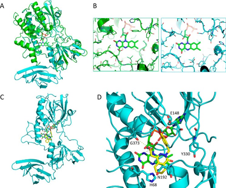
Figure 6.
Homology modeling of YUC2 and molecular docking of ponalrestat. A, structure of YUC2 (in cyan) based on homology modeling with FMO (in green). B, structural details of the binding pocket of FMO (in green) and YUC2 (in cyan) in complex with FAD. C, a complex structure of YUC2 and ponalrestat based on a docking simulation. D, structural details of the binding pocket of YUC2 in complex with ponalrestat and FAD. Dashed lines indicate hydrogen bonds.

