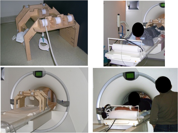
Fig. 1.
Self-built MRI compatible ergometer. Participants lay in prone position inside the MRI scanner. The ergometer was self-built and nonmagnetic (mainly built of wood). Moving of the work load was achieved via a pulley system. The left foot was secured to a padded foot loop. This loop was connected to a basket using a rope. Knee-extension led to an upward movement of the load. To ensure the correct placement of the thigh muscles on the magnetic coil, the thigh was secured to the coil using Velcro straps

