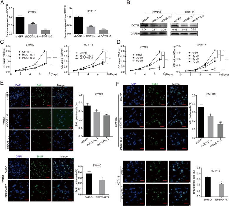
Fig. 4.
DOT1L silencing or inhibition blocks cell proliferation of CRC cells in vitro. a Relative mRNA expression of DOT1L detected by using qRT-PCR in SW480 and HCT116 colorectal cancer cell lines after DOT1L knockdown. shGFP vectors were used as control. b Protein expression of DOT1L detected by using Western blot in SW480 and HCT116 cells after DOT1L knockdown. Gray ratio of each blot was analyzed by using the Image J software and DOT1L/GAPDH ratio was shown. c, d Cell growth curve was determined by using MTT assay in SW480 and HCT116 cells after DOT1L knockdown or treatment with its specific inhibitor EPZ004777 with different concentrations for 1/3/5/7 days. e, f Cell proliferation was detected by using BrdU immunofluorescence in SW480 and HCT116 cells after DOT1L knockdown or inhibited by using EPZ004777 for 48 h (30 μM in SW480 and 50 μM in HCT116)

