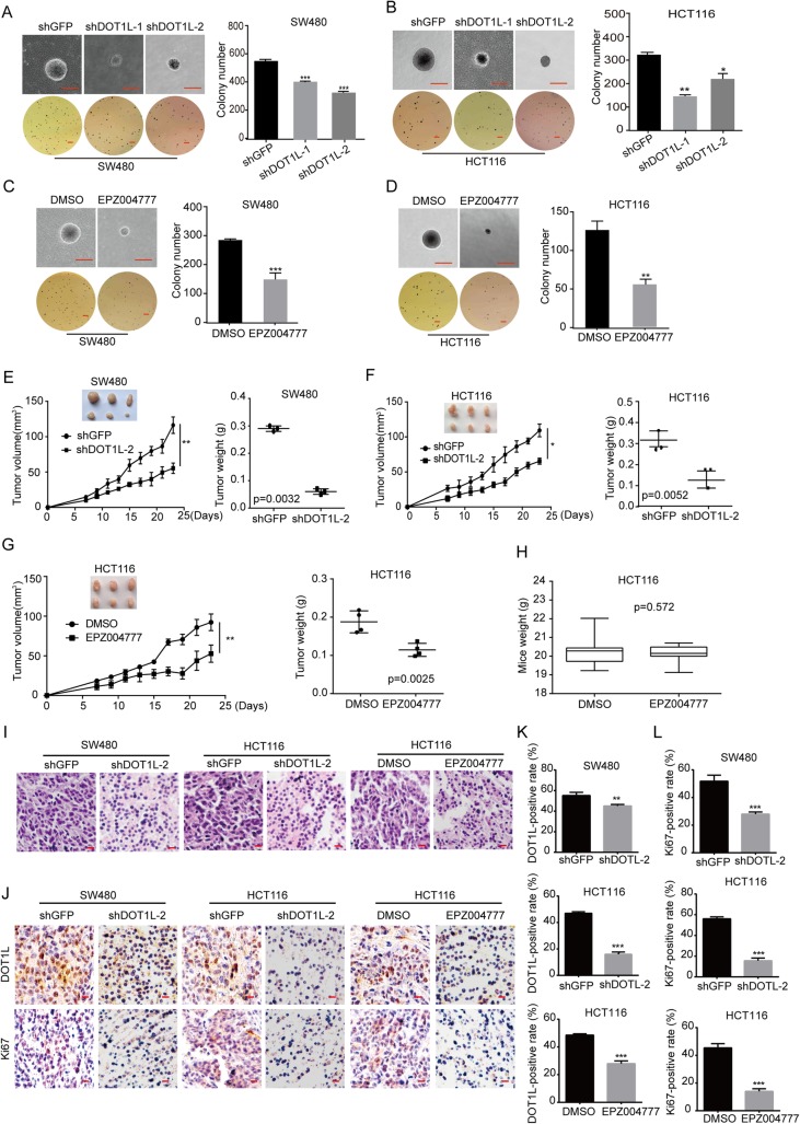
Fig. 5.
DOT1L silencing or inhibition suppresses tumorigenicity of CRC cells in vivo. a–d Self-renewal capacity detected by using soft agar assay in SW480 and HCT116 cells after DOT1L knockdown for 3 weeks or inhibited by using EPZ004777 for 2 weeks (30 μM in SW480 and 50 μM in HCT116). e, f The capacity of tumorigenicity was detected in BALB/c-nu mice subcutaneous injected with SW480 and HCT116 cells after DOT1L knockdown. Tumor volume was detected every 2 days from a week after subcutaneous injection. Tumor weight was measured after the tumors were removed from the bodies when the experiment was ended. g, h The capacity of tumorigenicity was detected in BALB/c-nu mice subcutaneous injected with HCT116 cells. After a week, mice were treated with EPZ004777 (100 mg/kg/day, diluted into PBS with 10% DMSO) or control solvent via intraperitoneal injection for 16 days. Tumor volume was detected every 2 days from the first time of intraperitoneal injection. Tumor weight and mice weight were measured after the tumors were removed from the bodies when the experiment was ended. i H&E staining of xenografts obtained from subcutaneous injecting SW480 and HCT116 cells after DOT1L knockdown or inhibition within mice. j–l IHC staining of DOT1L and Ki67 in xenografts obtained from subcutaneous injecting SW480 and HCT116 cells after DOT1L knockdown or inhibition within mice. Signal-positive rate was analyzed by using the IHC profiler in the Image J software

