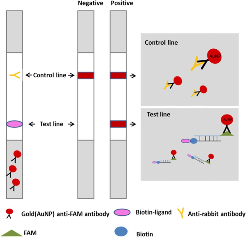
Fig. 1.

Detection of the RAA amplicons by lateral flow strip. The sample pad contains gold-labeled anti-FAM antibodies, the test line was coated with biotin-ligands, and control line was coated with anti-rabbit antibodies. The double-labeled amplicons (FAM and biotin) were diffused through the chromatographic membrane, and when they diffused to the test line, the products were captured by the biotin -ligands, resulting in an appearance of red-pink color. Non-captured particles will be fixed at the control line by anti-rabbit antibodies. In the absence of target amplicons, color will appear at control line only [26]
