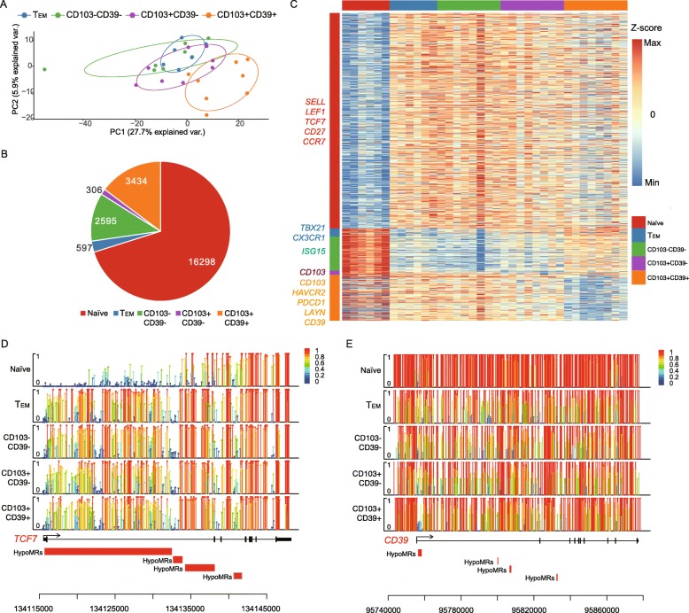
Fig. 2.
Whole-genome methylation profiling across multiple CD8+ T cell subtypes. a PCA analysis based on methylation profiles of CD8+ T cells in four T cell subtypes. b The graph shows the number of HypoMRs identified among five T cell subtypes. c Heat map showing the HypoMRs in the five subtypes. Color gradation from blue to red represents low to high DNA methylation levels. Selected genes associated with the HypoMRs were listed at the left side. d, e Lollipop plots for the nucleotide-resolution methylation level of the TCF7 and CD39 loci. Each covered cytosine is displayed as a bar with a large round head. The color and height of the bar indicate the methylation level

