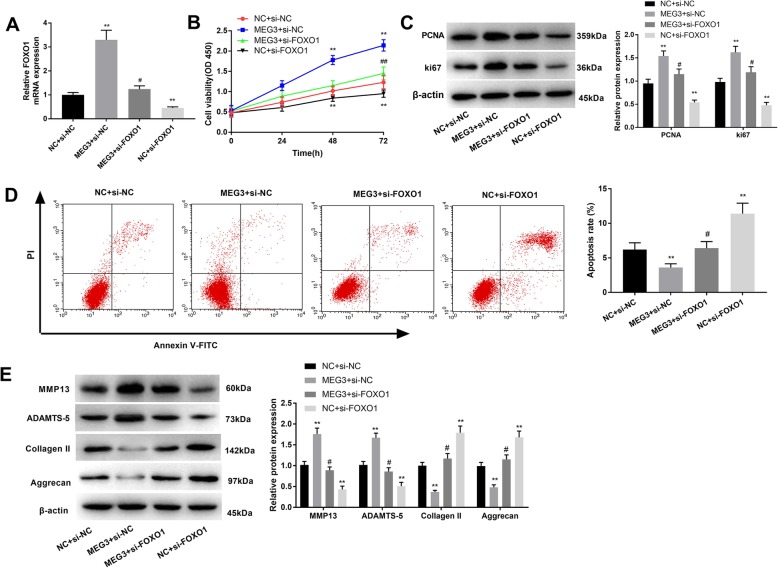
Fig. 5.
FOXO1 contributed to the impacts of MEG3 in OA. a, qRT-PCR showed the FOXO1 expression after diverse treatments. b and c, CCK-8 and western blot analyses were conducted to explore cell proliferation. d, cell apoptosis was determined using flow cytometry. e, the effects of FOXO1 on ECM degradation were examined by using western blotting. *, P < 0.05 compared with NC + si-NC. #, P < 0.05 compared with MEG3 + si-NC

