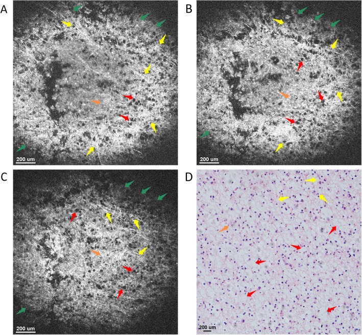
Fig. 8.
En face OCT images of high-grade glioma tissue ex vivo at imaging depths of (a) 150 μm, (b) 350 μm, and (c) 550 μm. The microstructures, e.g., mucin-like stroma (green arrows), glioblastoma multiforme (blue arrows), glioma (orange arrows), as well as fibrous (yellow arrows) and vesicles (red arrows) could be clearly identified with good correspondence to the histology. d Histology of glioma tissue image with hematoxylin and eosin: 100×

