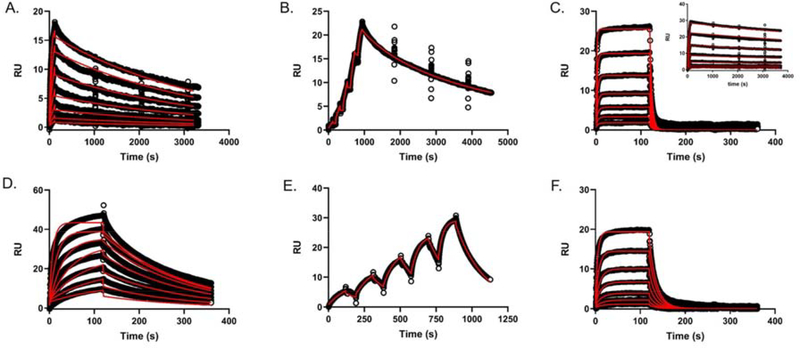
Figure 8. Surface Plasmon Resonance of Wild-Type and Mutant EapH1/H2 binding to immobilized hNE.
(A) EapH2 WT (dose-response kinetics with elongated dissociation phase, [EapH2] ranging from ~1.8x – 118x the EapH2 Ki), (B) EapH2 WT (single-cycle kinetics, [EapH2] ranging from ~3x – 47x the EapH2 Ki), (C) EapH1 L59F ([EapH1] ranging from` 0.3x – 20x the L59F Ki); inset: WT EapH1 dose-response sensorgram 3, (D) EapH2 T125A, ([EapH2] ranging from ~0.3x – 19x the T125A Ki), (E) EapH2 T125A, (single-cycle kinetics, [EapH2] ranging from ~0.3x – 5x the T125A Ki), (F) EapH2 N127A ([EapH2] ranging from ~ 0.2x – 10x the N127A Ki). All sensorgram series were evaluated with a Langmuir kinetic model as described in the materials and methods section.

