Figure 7.
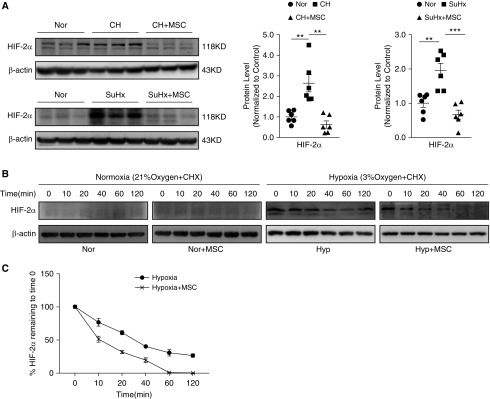
MSC-CM inhibited hypoxia-induced HIF-2α (hypoxia-inducible factor-2α) protein expression. (A) The protein expression of HIF-2α in lung tissues from CHPH (upper) and SuHx-PH (below) model with or without MSC treatment. Error bars represent means ± SEM (n = 6 in each group). **P < 0.01 and ***P < 0.001. (B) PMECs were exposed for 72 hours to normoxia/hypoxia with/without MSC-CM. Then cells were maintained in the presence of cycloheximide. The protein expression levels of HIF-2α in PMECs were measured by Western blot. (C) The trace of HIF-2α protein degradation (% HIF-2α protein remaining to time 0). Points, mean from three experiments. Error bars represent means ± SEM (n = 3 in each group).

