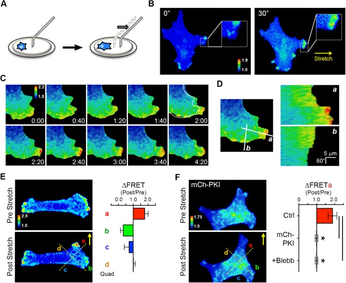
FIGURE 5:
PKA is activated upon acute mechanical stretch and is required for SKOV-3 cell durotaxis. (A) Schematic of technique using a glass microneedle to impart acute directional stretch on individual cells by deformation of the underlying hydrogel (see text for details). (B) An SKOV-3 cell expressing pmAKAR3, plated on a hydrogel coated with fibronectin for 4 h and then imaged by FRET microscopy, before and 30 s after application of mechanical stretch by a microneedle in the direction indicated by the arrow. Warmer colors correspond to higher PKA activity as assessed by FRET ratio. (C) Time course of FRET ratio indicating PKA activity in an SKOV-3 cell expressing pmAKAR3 before and after application of mechanical stretch. Immediately before the 2:00 min mark, stretch was applied directly to the right of the panel. Deformation of the cell is highlighted by the outline of the cell before the stretch overlaid at 2:00 min. (D) Line scan analyses in the direction of (a) and orthogonal to the axis of stretch (b) show a unique increase in PKA activity along the axis of stretch after stimulation. (E) FRET ratio images were sectioned into quadrants a through d, depicted by dotted lines, for quantification of directional response. The change in PKA activity before and after stretch (ΔFRET) of SKOV-3 cells expressing pmAKAR3 was calculated in quadrants proximal (quadrant a), orthogonal (quadrants b and d), or contralateral (quadrant c) to the stretch (mean ± SEM; n = 6). (F) Change in PKA activity before and after stretch (ΔFRET) in quadrant a of SKOV-3 cells coexpressing pmAKAR3 and mCh-PKI (shown), coexpressing pmAKAR3 and mCherry, or SKOV-3 cells expressing only pmAKAR3 but pretreated with 25 μM blebbistatin (Blebb) for 10 min (mean ± SEM; n = 9 or 5 cells for control or mCh-PKI cells, respectively; *p < 0.005 [Student’s t test]).

