FIGURE 6:
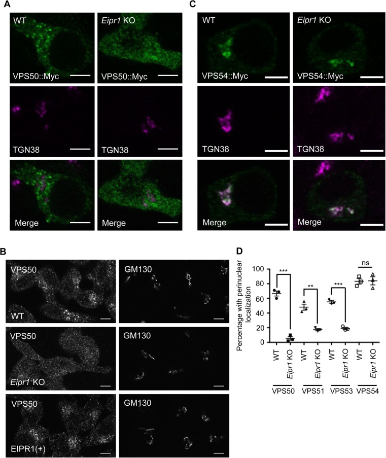
EIPR1 is required for the localization of EARP in insulin-secreting cells. (A) Representative images of 832/13 WT and Eipr1 KO cells transfected with VPS50::13Myc (VPS50::Myc) and costained with anti-Myc and anti-TGN38 antibodies. In WT cells, VPS50::Myc is localized to puncta but in Eipr1 KO cells, fluorescence is diffuse throughout the cytoplasm. The punctate pattern of localization of VPS50::Myc overlaps only partially with TGN38. Scale bars = 5 μm. (B) Representative images of 832/13 (WT), Eipr1 KO, and EIPR1(+) cells stained with anti-VPS50 antibody. Endogenous VPS50 is punctate in WT cells, but diffuse throughout the cytoplasm in Eipr1 KO cells. Scale bars = 5 μm. (C) Representative images of 832/13 cells (WT) and Eipr1 KO 832/13 (Eipr1 KO) cells transfected with VPS54::13Myc (VPS54::Myc) and costained with anti-Myc and anti-TGN38 antibodies. In both WT and Eipr1 KO cells, VPS54::Myc is localized to perinuclear puncta that largely overlap with TGN38. Scale bars = 5 μm. (D) Localization of Myc-tagged VPS50, VPS51, and VPS53, but not VPS54, is disrupted in Eipr1 KO cells. Shown is the percentage of cells with perinuclear and punctate staining of each indicated subunit. The experiment was repeated three times and ∼100 cells were imaged per experiment. Cells with very high expression level were not included in the counting because overexpression of the individual subunits leads to their mislocalization to the cytoplasm in WT cells. ***, p < 0.001; **, p < 0.01; ns, p > 0.05.

