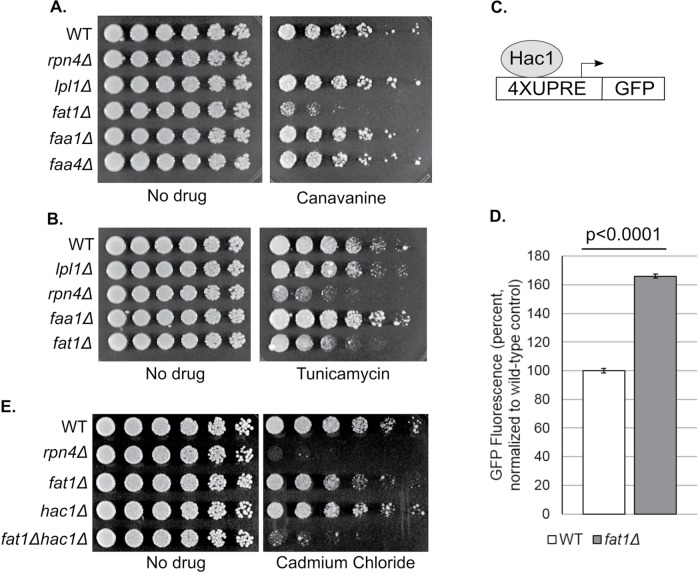
FIGURE 1:
VLCFA dysfunction leads to ER stress and compensatory induction of the UPR. (A) Growth of the indicated strains in the presence or absence of the amino acid analogue canavanine (2.5 μg/ml). Cells were spotted in threefold serial dilutions and cultured at 30°C for 2 d. (B) Growth of the indicated strains in the presence or absence of tunicamycin (2.5 μg/ml), an inducer of ER stress. Cells were spotted in threefold serial dilutions and cultured at 30°C for 2 d. (C) Schematic of the UPR reporter. Four copies of the Hac1 recognition sequence (UPRE) were fused to the coding region of GFP and integrated into the genome. (D) Constitutive induction of the UPR in the fat1Δ mutant. Results represent the mean GFP signal from four technical replicates and are normalized to the wild-type (WT) control. Error bars represent SDs. Results were also significant by two-tailed Student’s t test (p < 0.0001). (E) Abrogation of UPR induction by Hac1 sensitizes fat1Δ cells to ER stress. The indicated strains were cultured in the presence or absence of cadmium chloride (60 μM), a known inducer of the UPR (Gardarin et al., 2010). Cells were spotted in threefold serial dilutions and cultured at 30°C for 2–3 d.

