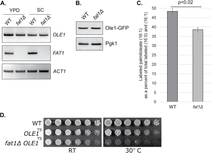
FIGURE 6:
Ole1 function is dependent on Fat1. (A) OLE1 mRNA levels, as determined by RT-PCR, in wild-type (WT) and fat1Δ strains grown in rich (YPD) or synthetic complete (SC) media. Middle, FAT1 mRNA levels; bottom, ACT1 mRNA levels (loading control). (B) Ole1 protein levels in the wild type and fat1Δ mutant. Whole-cell extracts were prepared at steady state from exponentially growing cultures and analyzed by SDS–PAGE followed by immunoblotting. An integrated Ole1-GFP strain was used to allow for immunohistochemical detection (top, anti-GFP antibody). Bottom, anti-Pgk1 antibody (loading control). (C) Generation of 13C-labeled palmitoleic acid (16:1) from 13C-labeled palmitic acid (16:0) in wild-type and fat1Δ cells. Cells were cultured with labeled palmitic acid at 50 μM for 7 h. Total lipids were extracted, and the abundance of 13C-labeled palmitoleic acid was determined by FT-ICR MS. Relative abundance of 13C-labeled palmitoleic acid is shown as a percent of the total labeled (16:0) and (16:1) fatty acid detected. Error bars represent SDs from two independent experiments. Results were also significant by two-tailed Student’s t test (p < 0.02). (D) Loss of Fat1 strongly compromises growth of a temperature-sensitive Ole1 mutant. Cells were spotted in threefold serial dilutions and cultured at 24°C (“RT”) and 30°C for 2–4 d.

