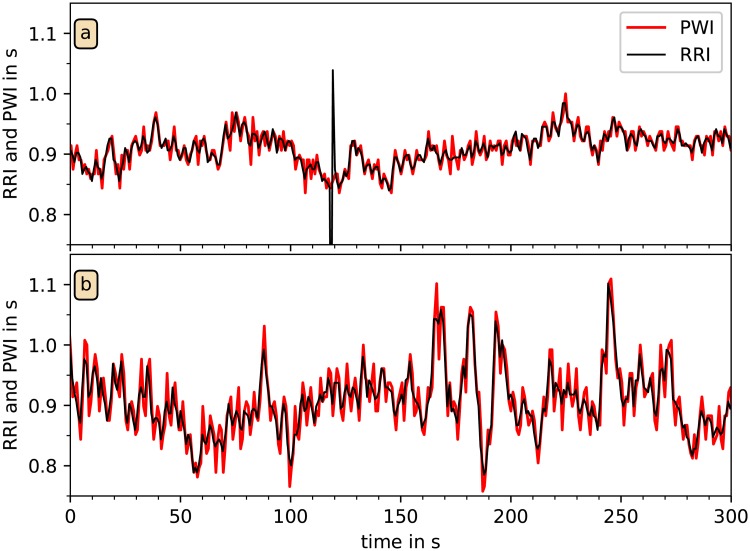
Fig 6. Comparison of tachograms from RRI and PWI.
In these two examples from different subjects, RRI derived from the ECG (black) and PWI independently derived from wrist accelerometry (red) are plotted versus time. All detected PWP and all R peaks were used; the PWI are strongly correlated with RRI. However, unexpected heartbeat events, as for example the premature beat at t = 120 s in (a), are not present in the PWI signal.

