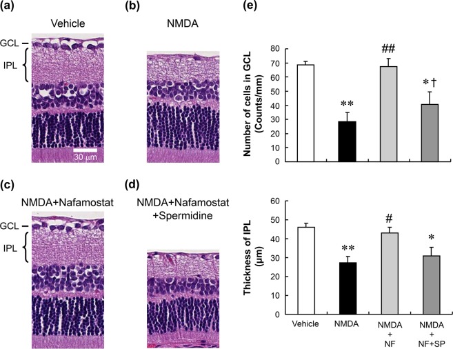
Figure 5.
Reversal by spermidine of the neuroprotective effect of nafamostat. Panels a–d show the typical histological appearance of rat retinas after intravitreal injections of vehicle (a), NMDA alone (b, 20 nmol/eye), NMDA plus nafamostat (c, 10 nmol/eye) and NMDA with nafamostat plus spermidine (d, 50 nmol/eye). The scale bar equals 30 µm. Panel e shows the changes in the GCL cell number (upper) and IPL thickness (lower) after intravitreal injections of vehicle (open column), NMDA alone (closed column, 20 nmol/eye), NMDA plus nafamostat (NF, light-grey column, 10 nmol/eye) and NMDA plus nafamostat and spermidine (SP, dark-grey column, 50 nmol/eye). Each value represents the mean ± S.E. for four to five rats. *P < 0.05; **P < 0.01, compared with vehicle, #P < 0.05; ##P < 0.01, compared with NMDA alone and †P < 0.05, compared with NMDA plus NF by Tukey’s multiple comparison test.

