Figure 1.
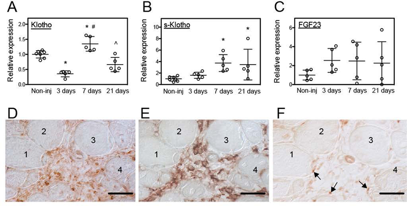
Acute injury affected klotho expression in skeletal muscle. (A) QPCR data showing reduced klotho expression 3-days after sterile muscle injury and increased expression 7-days after injury of tibialis anterior (TA) muscle of 5-months old wild-type (Wt) mice. (B) QPCR data showing that s-klotho expression in muscle increased at 7- and 21-days following injury. (C) QPCR data showing that Fgf23 expression was not significantly affected by muscle injury. N = 5 per data set. * indicates significantly different from non-injured muscle, # indicates significantly different from muscle 3-days post-injury and ^ indicates significantly different from muscle 7-days post-injury at P < 0.05 analyzed by one-way ANOVA. Bars = standard deviation. (D-F) Serial sections of TA muscles of 5-months old mice at 7-days post-injury that were immunolabeled for Klotho (D), CD206 (E) or Pax7 (F). An inflammatory lesion surrounded by four muscle fibers (1–4) shows highest levels of Klotho (D; reddish-brown), elevated numbers of CD206+ macrophages (E; reddish brown) and Pax7+ cells associated with the neighboring muscle fibers (arrows) (F). Bars = 50 μm.

