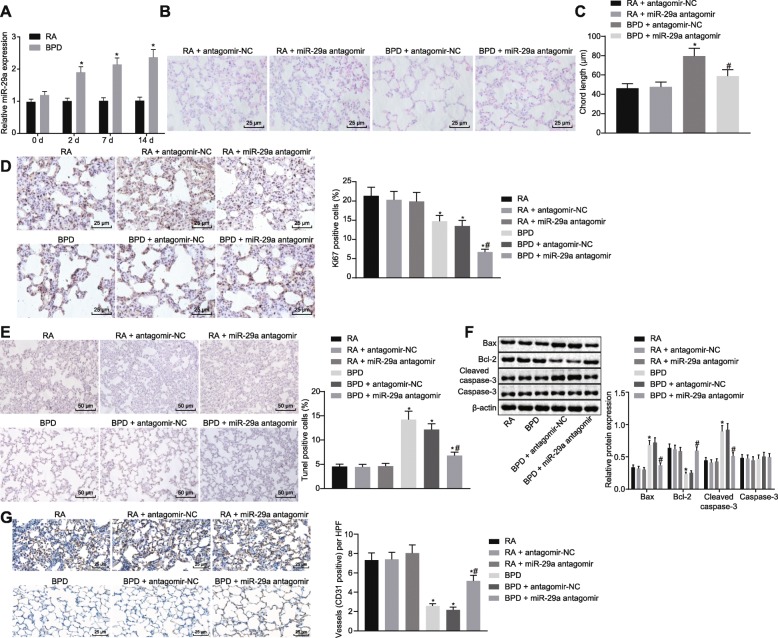
Fig. 2.
Inhibition of miR-29a impedes hyperoxia-induced BPD in neonatal mice. A, The expression of miR-29a in lung tissues of neonatal mice under exposure of RA or hyperoxia determined by RT-qPCR. * p < 0.05 vs. neonatal mice under exposure of RA (the RA group). B, HE staining analysis of lung tissues in each group (× 400). C, Alveolar chord length of lung tissues in each group. D, Positive expression rate of Ki67 protein in lung tissues of each group determined by immunohistochemistry (× 400). E, Cell apoptosis in lung tissues of each group assessed by TUNEL staining (× 200). F, Western blot analysis of apoptosis-related proteins (Bax, Caspase-3 and Bcl-2) in lung tissues of each group. G, Positive expression rate of CD31 protein in lung tissues of each group determined by immunohistochemistry (× 400). In panel C-F, * p < 0.05 vs. neonatal mice under exposure of RA (the RA group). # p < 0.05 vs. neonatal mice with hyperoxia-induced BPD receiving antagomir-NC (the BPD + antagomir-NC group). N = 15. Measurement data were expressed as mean ± standard error. Data comparison between two groups was examined by unpaired t-test. miR-29a, microRNA-29a; BPD, bronchopulmonary dysplasia; RA, room air; Bax, Bcl-2-associated X protein; Bcl-2, B-cell lymphoma 2; RT-qPCR, reverse-transcription quantitative polymerase chain reaction; HE, hematoxylin-eosin; RAC, radial alveolar counts; TUNEL, TdT-mediated dUTP-biotin nick end-labeling; NC, negative control

