Fig. 3.
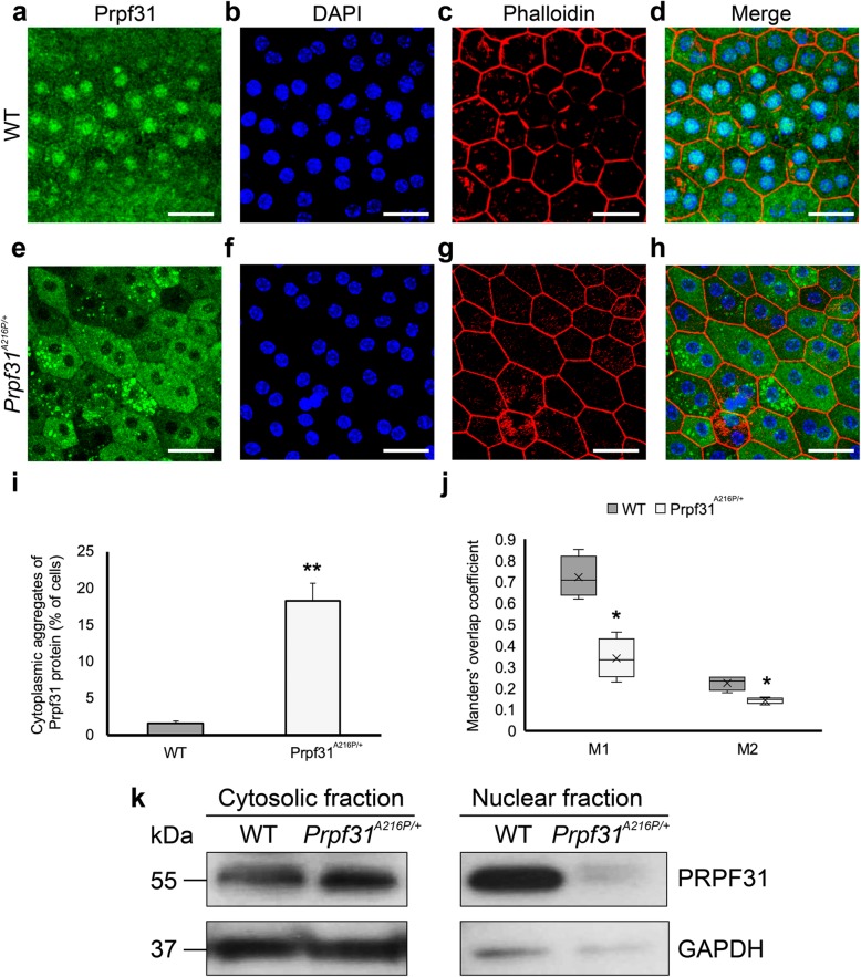
Large cytoplasmic aggregates of Prpf31 protein were observed in the RPE of Prpf31A216P/+ mice. Whole-mount of the RPE layer from WT (a-d) and Prpf31A216P/+ mice (e-h) were immunostained with anti-Prpf31 antibodies (a, e). Cell nuclei were stained with DAPI (b, f), and TRITC-phalloidin was used to visualize the F-actin microfilaments (c, g). Prpf31 signal was mainly localized in the nuclei of WT RPE cells (a), while large protein aggregates stained for PRPF31 were observed in the cytoplasm of RPE cells of mutant Prpf31A216P/+ mice (e). The bars in graph i represent the percentage of RPE cells with cytoplasmic aggregates of Prpf31 protein ± SEM in WT and Prpf31A216P/+ whole mount RPE samples (n = 1200 cells were counted from 4 mice in each group). The boxplot j represents the Manders’ overlap coefficient of DAPI+Prpf31/DAPI colocalization (M1) and Prpf31 + DAPI/Prpf31 colocalization (M2) in WT and Prpf31A216P/+ whole-mount RPE samples (n = 4 in each group). Expression of Prpf31 protein was evaluated in the cytosolic and nuclear fractions by Western blot (k). Statistically significant differences were determined by t-test or Mann-Whitney U-test (*p < 0.01, **p < 0.001). Scale bars represent 25 μm

