Fig. 7.
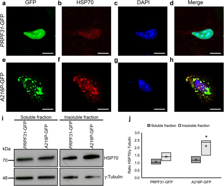
Overexpression of A216P-GFP induces HSP70 activation in ARPE-19 cells. Immunostaining of cultured ARPE-19 cells transfected with PRPF31-GFP (a-d) orA216P-GFP (e-h) displays PRPF31 aggregation in the cytoplasm of the cells overexpressing A216P-GFP (green) and colocalization of HSP70 (red) in the aggregates (e-h). Images correspond to a maximum projection of a Z-stack. Western blot analysis (i) and densitometry quantification (j) of the soluble and insoluble fraction of the transfected cells showing an increment of HSP70 concentration in the detergent-insoluble fraction of the cells transfected with A216P-GFP. Anti-γ-tubulin antibody was used as loading control (i). The boxplot j represents the ratio HSP70/γ-tubulin in soluble and insoluble fraction of PRPF31-GFP and A216P-GFP transfected ARPE-19 cells (n = 3 in each group). Statistically significant differences were determined by Mann-Whitney U-test (*p < 0.05). Scale bars represent 10 μm

