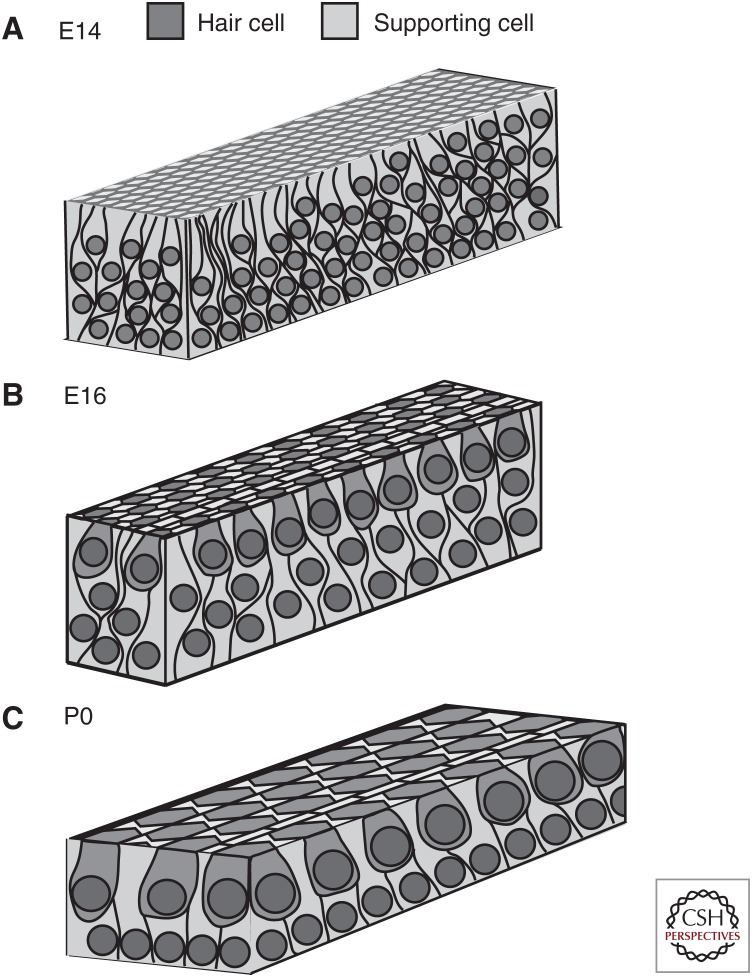
Figure 1.
Changes in distribution of prosensory cells during cochlear outgrowth. Each drawing represents a region near the midbase of the cochlea. (A) At E14, the epithelium is highly pseudostratified. There is no cellular organization and no morphological differentiation has occurred. (B) At E16, immature hair cells have begun to become organized into rows and stratification has decreased although more than two layers of cells are still present. Note the overall decrease in cell density as a result of cellular migration and extension. (C) By P0, the mature cellular pattern is present with a luminal layer of hair cells resting on a basal layer of supporting cells.

