Figure 3.
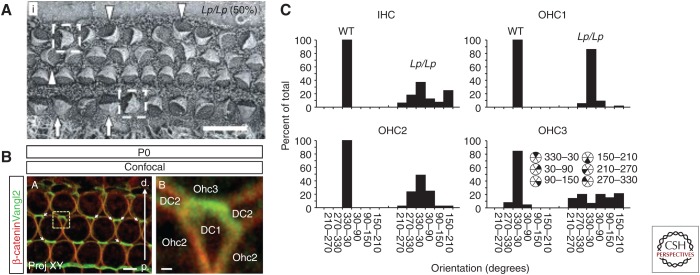
Vangl2 regulates stereociliary bundle orientation in the cochlea. (A) Scanning electron micrograph of the organ of Corti from a Vangl2Lp/Lp mouse at E18.5. Stereociliary bundles show varying degrees of misorientation (indicated by arrowheads, arrows, and boxed cells). (B) Confocal image of the surface of the outer hair cell (OHC) region from the organ of Corti of a P0 mouse. Vangl2 is localized to the junctions between the distal sides of Deiters cells (DCs) and the proximal sides of OHCs. (C) Frequency histograms illustrating the distribution of bundle orientations, based on cell type, in control and Vangl2Lp/Lp cochleae at E18.5. Note that although bundles in wild-type (WT) animals show a limited distribution centered around the optimal (0°) orientation, in Vangl2Lp/Lp bundles show varying degrees of misorientation based on location. Scale bars, 10 μm (A); 4 μm (B, left); 0.5 μm (B, right). IHC, Inner hair cell. (Panels A and C from Montcouquiol et al. 2003; reproduced, with permission, from Springer Nature. Panel B from Giese et al. 2012; reproduced, with permission, from the authors.)

