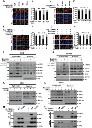
Fig. 4. CHK2-induced FOXK translocation from nucleus to cytoplasm in response to DNA damage is dependent on FOXK phosphorylation (FOXK1 S130 and FOXK2 S61).

(A) HEPG2 cells were transiently transfected with HA-FOXK2 WT or HA-FOXK2 S61A plasmid. Twenty-four hours after transfection, cells were treated with or without 20 μM cisplatin (CDDP). Representative images are shown. Scale bar, 10 μm. DAPI, 4′,6-diamidino-2-phenylindole. (B) Quantification of at least 100 cells from (A) viewed in five to eight random fields from n = 3 independent experiments. N: nucleus; C: cytoplasm. (C) HEPG2 cells were transiently transfected with HA-FOXK1 WT or HA-FOXK1 S130A plasmid. Twenty-four hours after transfection, cells were treated with or without 20 μM cisplatin (CDDP). Representative images are shown. Scale bar, 10 μm. (D) Quantification of at least 100 cells from (C) viewed in five to eight random fields from n = 3 independent experiments. (E to H) HA-FOXK2 WT (E) or HA-FOXK1 WT (G) plasmid was transfected into HEPG2 control cells or cells depleted CHK2. Twenty-four hours after transfection, cells were treated with or without 20 μM cisplatin (CDDP). Representative images are shown. Scale bar, 10 μm. Quantification of at least 100 cells from (E), (F), (G), or (H) viewed in five to eight random fields from n = 3 independent experiments is shown. (I and J) Western blot analysis was performed to assess endogenous FOXK cellular localization in A549 cells (I) or HEPG2 cells (J) transfected with control or CHK2 shRNA and treated with vehicle or 20 μM cisplatin for 24 hours. (K and L) Western blot analysis was performed to assess endogenous FOXK cellular localization in A549 (K) or HEPG2 (L) cells treated with CHK2 inhibitors and/or 20 μM cisplatin for 24 hours. (M and N) HEK293T cells transfected with HA-FOXK1 WT or HA-FOXK1 S130A (M) or HA-FOXK2 WT or HA-FOXK2 S61A (N) were treated with vehicle or 20 μM cisplatin for 24 hours and purified using anti–HA-agarose beads. The immunoprecipitates were then blotted with the indicated antibodies.
