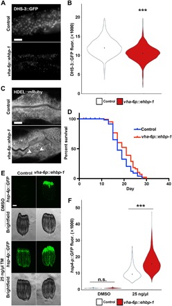
Fig. 3. Intestinal ehbp-1 overexpression is sufficient to drive lipid depletion, ER remodeling, and life-span extension.

(A) Representative compound micrographs of intestinal LD (via dhs-3p::DHS-3::GFP) in day 2 adult control and vha-6p::ehbp-1 animals. Scale bar, 10 μm. (B) Quantification of LDs (via DHS-3::GFP fluorescence) from (A). ***P < 0.001; using nonparametric Mann-Whitney test against age-matched control on corresponding RNAi. Plots are representative of three biological replicates and n = 2560 for control and n = 324 for vha-6p::ehbp-1. (C) Representative micrographs of ER morphology (mRuby::HDEL) in the intestine of control and vha-6p::ehbp-1 animals. ER remodeling is depicted by arrowheads. (D) Life-span data for wild-type N2 animals or vha-6p::ehbp-1 animals grown on empty vector RNAi from hatch. See table S1 for life-span statistics. (E) Fluorescent light micrographs of hsp-4p::GFP UPRER reporter animals (control and vha-6p::ehbp-1) treated with DMSO or tunicamycin (TM; 25 ng/μl) at L4 for 4 hours and then imaged as day 1 adults. Note that myo-2p::GFP was used as a coinjection marker, and high GFP signal in the pharynx is not an artifact or cause of EHBP-1 overexpression. Scale bar, 100 μm. (F) Quantification of hsp-4p::GFP fluorescence in control (white) and vha-6p::ehbp-1 (red) nematodes on DMSO or tunicamycin (25 ng/μl), using a BioSorter, following various RNAi treatments. The pharynx signal from the myo-2p::GFP coinjection marker was removed from quantification during analysis. ***P < 0.001; n.s., P > 0.10; using Mann-Whitney test. Data are representative of three independent trials with n = 172 to 3205 per strain.
