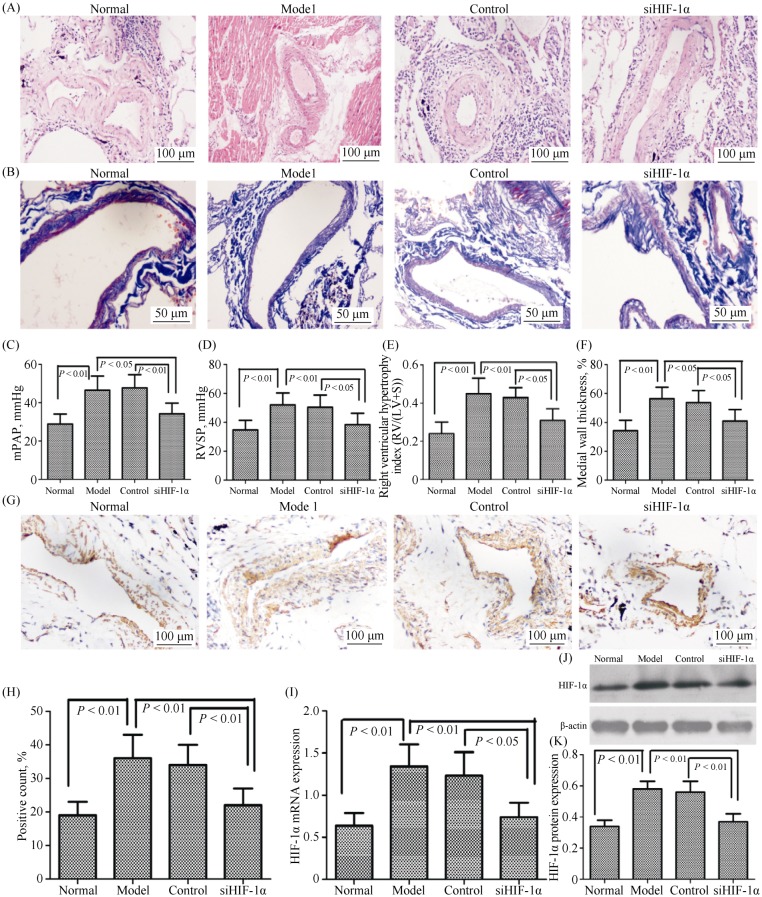
Figure 1. HIF-1α regulates the pathogenesis of hypoxic pulmonary hypertension.
(A): Representative images of hematoxylin-eosin staining (H & E) in lung tissues lesions, magnification 400 × (n = 6). Ten pulmonary arterioles were randomly examined for the structural integrity from hematoxylin-eosin staining images of lung tissues lesions by Image J software; (B): representative images of Masson staining in lung tissues lesions, magnification 400 ×; (C–D): right heart catheterization analysis of mPAP and RVSP on the surviving rats; (E): the degree of right ventricular hypertrophy expressed as RV/(LV + S); (F): the measurement of medial wall thickness of distal pulmonary vessels; (G–H): lung tissue sections were stained for HIF-1α (magnification, 200 ×); and (J–L): immunoblots of HIF-1α using pulmonary arteries isolated from the rats exposed to either normoxia or hypoxia. siHIF-1α, lentiviral vector-mediated short-hairpin RNA targeting HIF-1α; Control, lentiviral vector containing scramble siRNA. HIF-1α: hypoxia-inducible factor-1α; LV: left ventricle; mPAP: mean pulmonary arterial pressure; RV: right ventricle; RVSP: right ventricular systolic pressure; S: septum; siHIF-1α: small interfering hypoxia-inducible factor-1α; siRNA: small interfering RNA.

