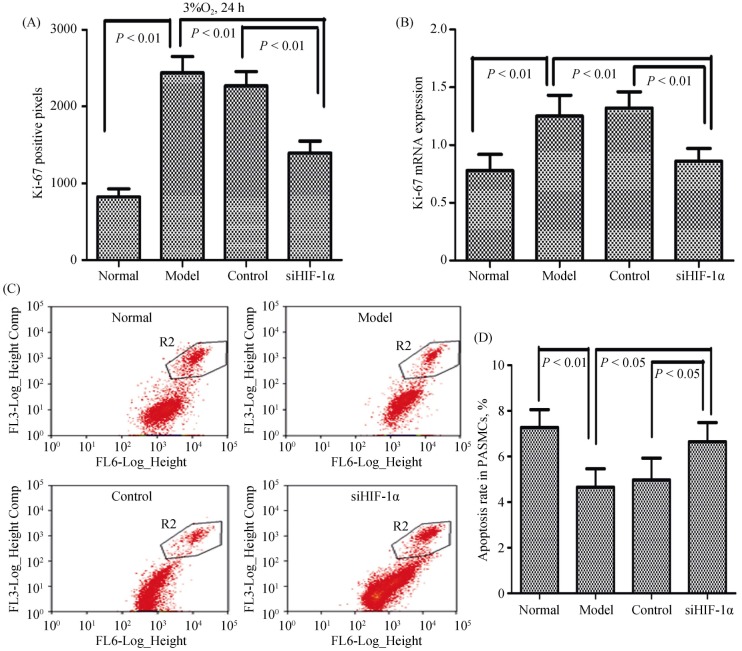
Figure 2. HIF-1α is involved in hypoxia-induced proliferation and apoptosis of PASMCs.
PASMCs were infected with lentiviruses harboring HIF-1α siRNA or scramble siRNA, and exposed to hypoxia (3% O2) for 24 hours. (A & B): The number of proliferation cells was determined by Ki-67 assay in cell number counting and expression of Ki-67 mRNA in the qRT-PCR; and (C–D): the cells were fixed and stained with propidium iodide, and cell cycle was analyzed by flow cytometry. A set of representative flow cytometry results (C), and statistical analysis of three independent experiments (D). HIF-1α: hypoxia-inducible factor-1α; PASMCs: pulmonary arterial smooth muscle cells; qRT-PCR: quantitative real-time polymerase chain reaction; siHIF-1α: small interfering hypoxia-inducible factor-1α; siRNA: small interfering RNA.

