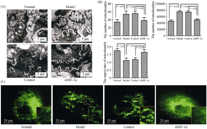
Figure 3. The morphology of mitochondria under hypoxia is dependent on HIF-1α in vivo and in vitro.
(A): Representative electron micrograph of lung tissues lesions in different groups (magnification, 10000 ×); (B): graphs indicate the mitochondria number, per area and average mitochondrial size; and (C): electron micrographs of mitochondria in PASMCs (magnification, 1000 ×). The normal group displayed elongated mitochondria, whereas the hypoxia model group displayed short or spherical shaped mitochondria. The administration of siHIF-1α markedly attenuated mitochondrial fragmentation in the PASMCs of hypoxia. HIF-1α: hypoxia-inducible factor-1α; PASMCs: pulmonary arterial smooth muscle cells; siHIF-1α: small interfering hypoxia-inducible factor-1α.

