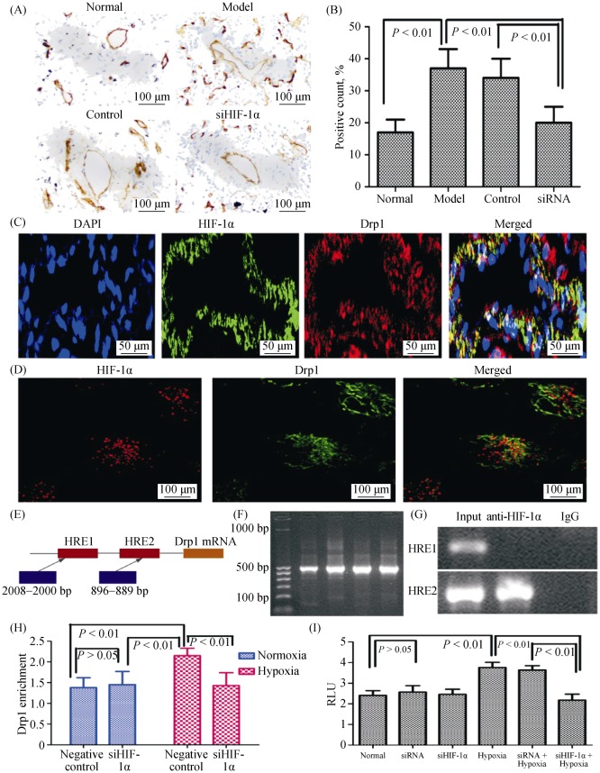
Figure 6. HIF-1α regulates mitochondrial function by Drp1 under hypoxia.
(A–B): Immunohistochemical evaluation of the expression of Drp1 in lung. Drp1 expression was increased in pulmonary vessels with hypoxia (magnification, 200 ×); (C): the sections were stained with Drp1 antibody (red) and co-stained with HIF-1α antibodies to demonstrate adventitia (green), DAPI to demonstrate cell nuclear (blue), respectively. Immuno-reactive Drp1 and HIF-1α were expressed both in the pulmonary vascular media and in the intima, and more intensively in the smooth muscle layer (magnification, 400 ×); (D): the PASMCs were stained with Drp1 antibody (red) and co-stained with HIF-1α antibodies to demonstrate adventitia (green), respectively. Immunoreactive Drp1 and HIF-1α were expressed both in PASMCs of mitochondrial under hypoxic conditions (magnification, 400 ×); (E): schematic diagram showing the hypoxic reactive elements in the promoter region of Drp1; (F–G): the levels of Drp1 promoter with HIF-1α in PASMCs were detected by chromatin immunoprecipitation assay; (H): the transcriptional activity of Drp1 promoter under hypoxia was detected by luciferase reporter gene; and (I): the effect of endogenous HIF-1 alpha expression on the activity of Drp1 promoter in siRNA PASMCs. DAPI: 4,6-diamond-2-phenyl insole; Drp1: dynamin-related protein 1; HIF-1α: hypoxia-inducible factor-1α; PASMCs: pulmonary arterial smooth muscle cells; siHIF-1α: small interfering hypoxia-inducible factor-1α; siRNA: small interfering RNA.

