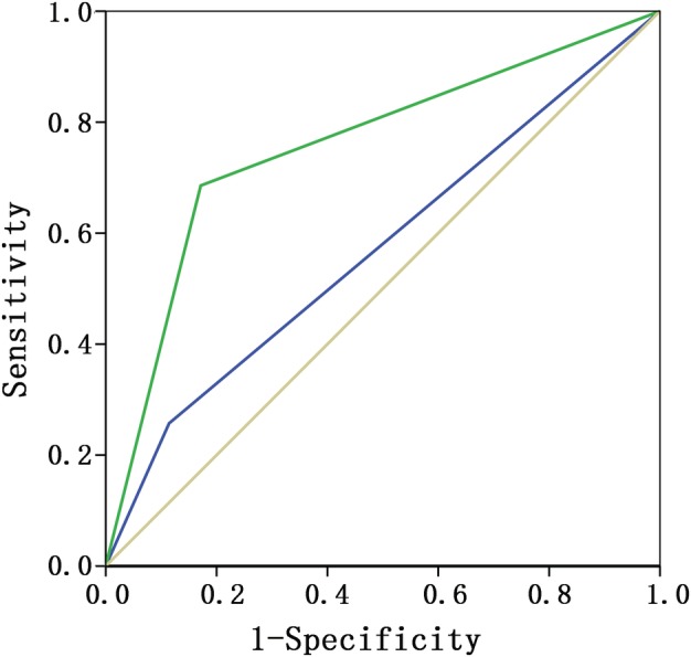
Figure 3.

The area under the ROC curve by PE and US of patients whose ALN was diagnosed as regular target annular lymph node with a peripheral cortical thickness ≥3 mm or eccentric target annular lymph node with a local cortical thickness ≥3 mm. ROC curves ( ) PE, (
 ) US and (
 ) reference.
