Figure 4.

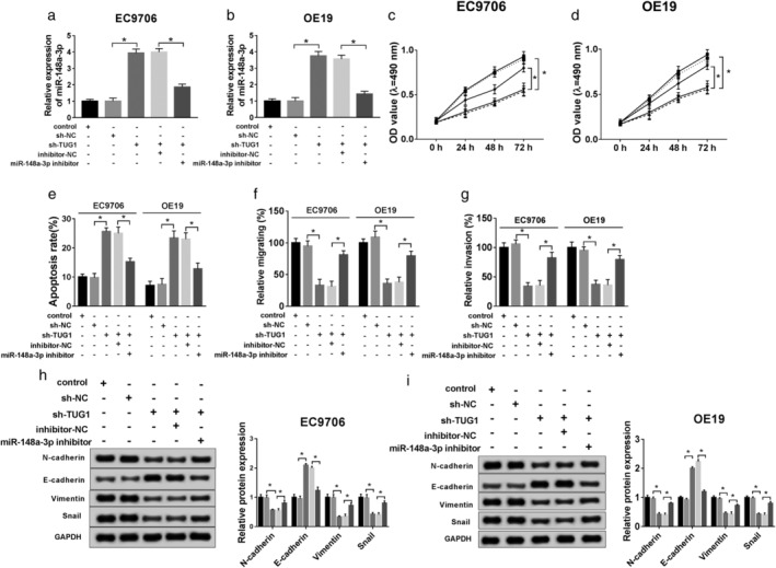
Knockdown of miR‐148a‐3p inverted functional effects from TUG1 deletion in ESCC in vitro. sh‐NC, sh‐TUG1, sh‐TUG1 + inhibitor‐NC, sh‐TUG1 + miR‐148a‐3p inhibitor was transfected into ESCC cells for subsequent experiment, separately. (a,b) QRT‐PCR was used to confirm the level of miR‐148a‐3p. (c,d) Cell viability was analyzed by MTT assay at stated times (0 hours, 24 hours, 48 hours, 72 hours) in EC9706 and OE19 cells. ( ) Control, (
 ) sh‐NC, (
 ) sh‐TUG1, (
 ) sh‐TUG1 + inhibitor‐NC, and (
 ) sh‐TUG1 + miR‐148a‐3p inhibitor. (e) Flow cytometry was conducted to analyze cell apoptosis rate. (f,g) Cell migration and invasion were evaluated by transwell assay. (h,i) The EMT‐related protein levels of N‐cadherin, E‐cadherin, Vimentin, and Snail were explored by western blot. *P < 0.05.
