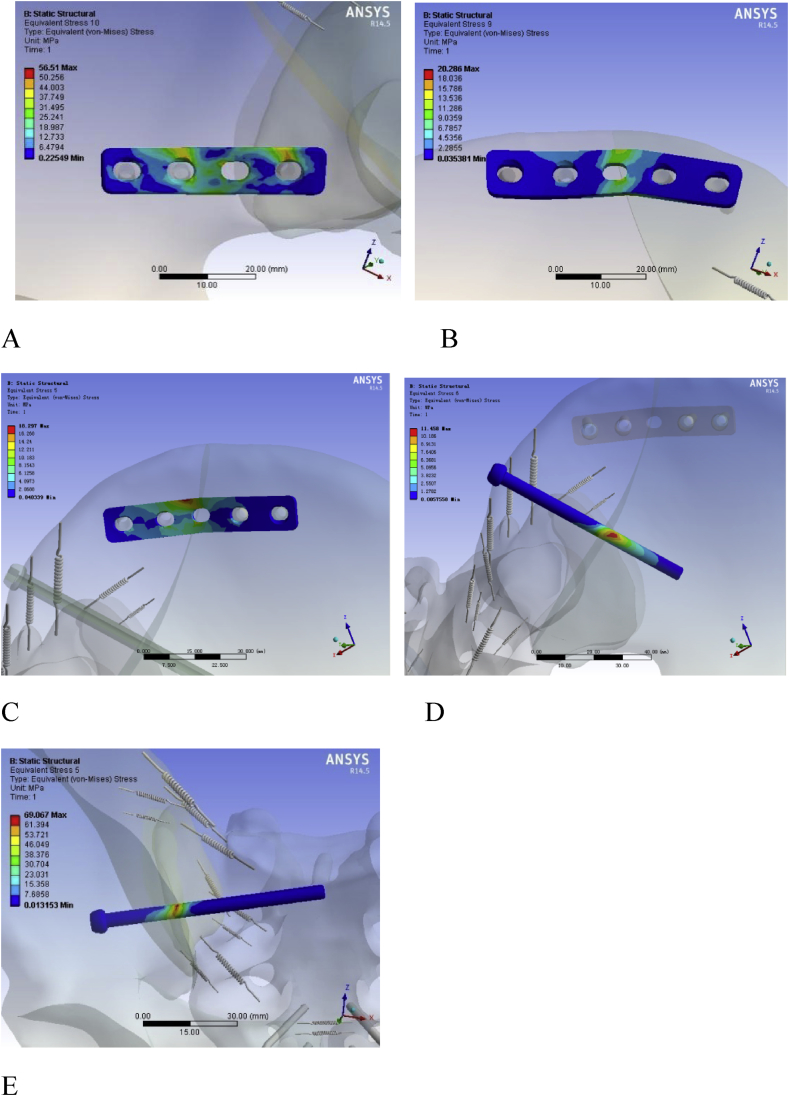
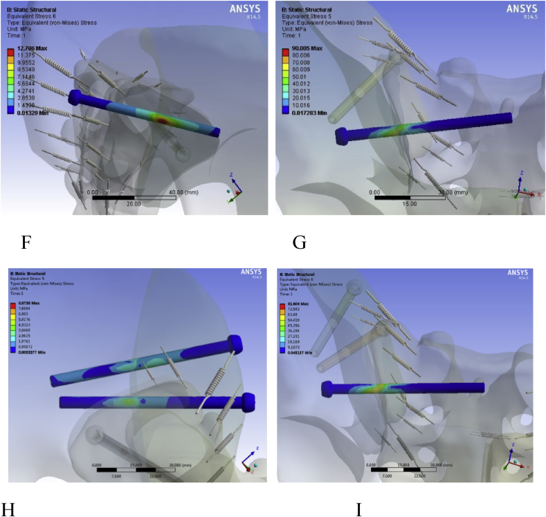
Figure 7.
Stress analysis of the fixation in the five models. A:Distribution diagram of plate stress fixed sacroiliac joint in model A; B: Distribution diagram of plate stress fixed crescent fragment in model A; C: Distribution diagram of plate stress fixed crescent fragment in model B; D: Distribution diagram of canulated screw stress fixed crescent fragment in model B; E: Distribution diagram of canulated screw stress fixed sacroiliac joint in model C; F: Distribution diagram of canulated screw stress fixed crescent fragment in model D; G: Distribution diagram of canulated screw stress fixed sacroiliac joint in model D; H:Distribution diagram of the two canulated screw stress fixed crescent fragment in model E; I:Distribution diagram of canulated screw stress fixed sacroiliac joint in model E. Whether it is the fixation of the crescent fragment or the SI joint, the cannulated screw can withstand more stress and is more evenly distributed than the plate, and the two cannulated screws are more advantageous.


