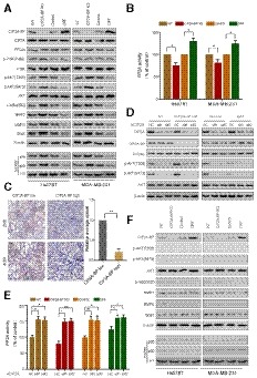
The expression levels of the indicated proteins were detected by immunoblotting analysis in CIP2A‐BP KO, LINC00665 ORF overexpressed (OE) and respective controls of TNBC cells.
PP2A activity was measured using a protein phosphatase assay kit. PP2A activity assay was performed to evaluate PP2A activity in CIP2A‐BP KO, LINC00665 ORF overexpressed (OE) and respective control of TNBC cells.
Immunohistochemistry (IHC) staining of p‐AKT from TNBC primary tumor (n = 20) with high or low micropeptide CIP2A‐BP expression. Lower panels show higher‐magnification images of insets in upper panels. Right panel is the quantification of IHC staining p‐AKT.
MDA‐MB‐231 cells were transfected with anti‐CIP2A siRNAs. Whole‐cell lysates of these cells were subjected to immunoblot analysis with CIP2A, CIP2A‐BP, PP2Ac, p‐AKT, AKT, and β‐actin antibodies.
CIP2A‐BP KO, LINC00665 ORF overexpressed (OE) and respective control MDA‐MB‐231 cells were transfected with anti‐CIP2A siRNAs. PP2A activity assay was performed to evaluate PP2A activity in the indicated cells.
The indicated cells were pretreated with specific antagonist against AKT (MK‐2206, 3 μM) for 1 h. Then, whole lysates of these cells were subjected to immunoblot analysis.
Data information: Data are representative of three independent experiments (B and E). Data were assessed by paired Student's
t‐test (B, C, and E) and are represented as mean ± SD. *
P < 0.05; **
P < 0.01. Scale bars: 50 μm (C).

