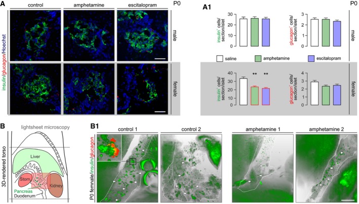
Figure 5. Both amphetamine and escitalopram reduce insulin immunoreactivity in female offspring at birth.

- Histochemical examples of neonatal pancreata used for the simultaneous detection of insulin and glucagon. Hoechst 33342 was used as nuclear counterstain. Scale bars = 80 μm. (A1) Quantitative data from n > 6 mice/sex from independent pregnancies. Note that insulin immunoreactivity was significantly reduced in the pancreas of female but not male offspring. Data were expressed as means ± SEM, **P < 0.01 (pair‐wise comparisons after one‐way ANOVA). Figure EV4C and C1 is referred to data on Slc6a4 −/− mice.
- Longitudinal cross‐sectional rendering of the neonatal mouse torso to aid pancreas visualization by light‐sheet microscopy. The area with semi‐transparent red overly is shown in B1. (B1) Insulin‐labeled pancreata (encircled by dashed lines) from respective pairs of control and amphetamine‐treated neonatal mice. Note the reduced size and labeling intensity for insulin of pancreatic islet‐like structures (arrowheads) upon intrauterine amphetamine exposure. Inset reveals that the core–shell cytoarchitecture of an islet is retained in optically cleared intact tissues simultaneously processed for the detection of insulin and glucagon. Many peripheral organs showed significant autofluorescence. Scale bar = 1.5 mm. Videos of orthogonally reconstructed tissues are available as part of the Appendix file.
