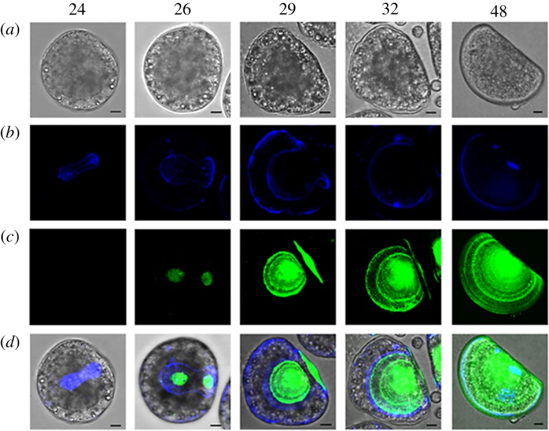
Figure 1.

Confocal images showing the time course of early shell formation in M. galloprovincialis from the trochophora (24 hpf) to the D-veliger stage (48 hpf) (lateral view). (a) Brightfield image of the embryo; (b) calcofluor fluorescent signal (blue), corresponding to the organic matrix; (c) calcein fluorescence signal (green), corresponding to CaCO3 deposition; (d) merged calcofluor, calcein and brightfield images. Scale bars, 10 µm. The secretion of the organic matrix is visible from 24 hpf (blue) followed by calcification (green) at 26 hpf, with a progressive expansion of the organic matrix and deposition of the shell starting from the central part of each valve. At 29 hpf the calcified valves are well developed onto the organic matrix. By 32 hpf calcification reaches the external margins of the organic matrix and expands towards the hinge region. At 48 hpf the whole shell is calcified and completely encloses the larval body. (Online version in colour.)
