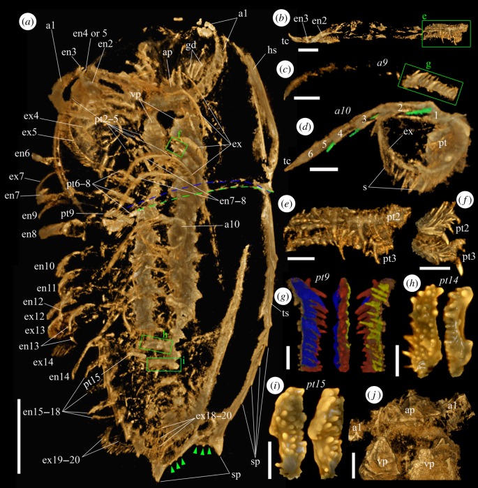
Figure 2.
Micro-CT images of an adult specimen (YKLP 11409) of Naraoia spinosa from the Chengjiang Lagerstätte. (a) Ventral view of the animal. Green dashed line marks the position of the posterior margin of the head shield, blue dashed line for the anterior margin of the trunk shield. Green arrowheads indicate tiny spines on the posterior margin of the trunk shield. (b) Right second and third appendages, exopods not preserved. (c) Right ninth appendage, exopod not preserved. (d) Left tenth appendage. Green coloration highlights apical setae on endopodal podomeres 1–4. (e) Protopods of right second and third appendages, interior view (green rectangle in (b)). (f) Protopods of left second and third appendages, interior view (one of the green rectangles in (a), viewing from the back side). (g) Protopod of right ninth appendage (green rectangles in (c)) in posterior, interior and anterior views. Blue, red and yellow colorations highlight different rows of spines. (h) Protopod of left 14th appendage in ventral and posteroventral views. (i) Protopod of left 15th appendage in ventral and anteroventral views. (j) Hypostomal complex and basal parts of first appendages (antennae), ventral view. Scale bars, 5 mm for (a), 1 mm for (b–d, j) and 500 µm for (e–i). Abbreviations as in figure 1. Numerals 1–6 indicate endopodal podomeres. (Online version in colour.)

