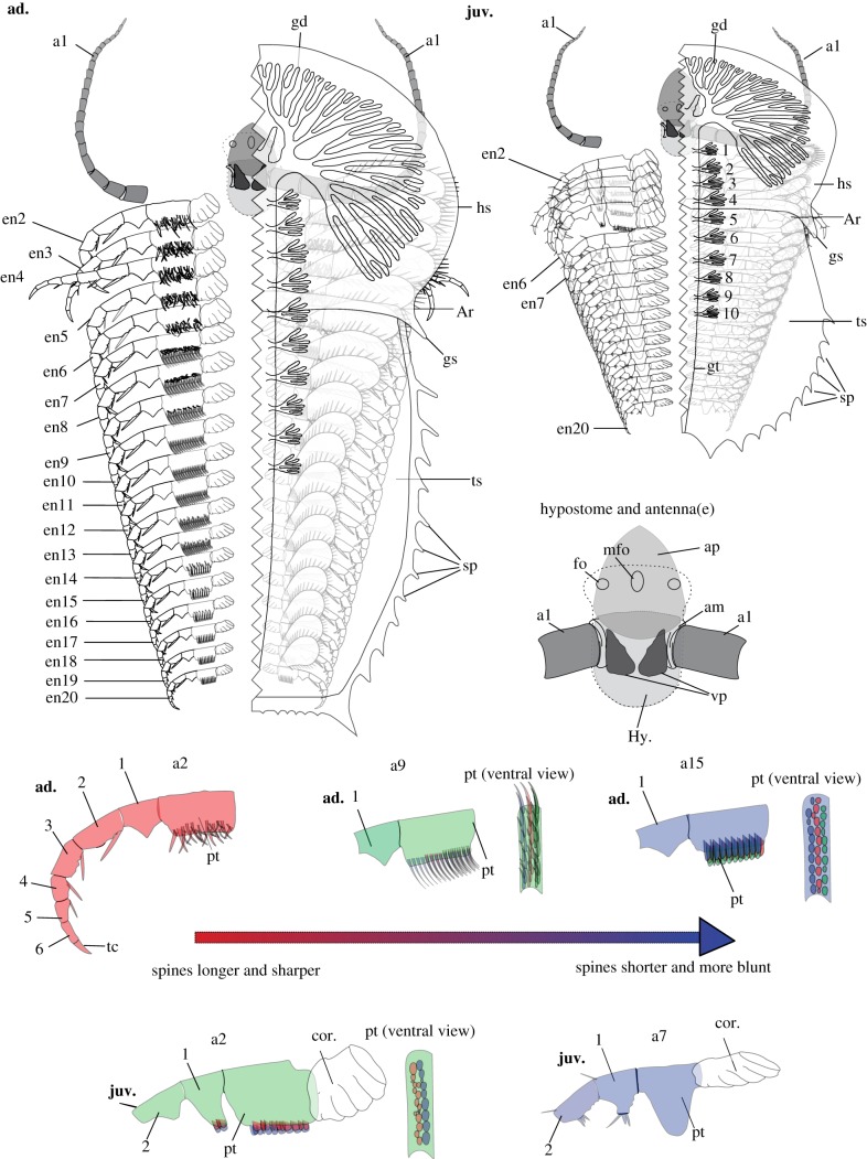
Figure 3.
Line drawings showing differentiation of the appendages of Naraoia spinosa and the difference between juvenile and adult. Left- hand upper: a detailed line drawing of N. spinosa, of early-adult stage of growth (ad.), based on YKLP 11409. Right- hand upper: a detailed line drawing of N. spinosa, of mid-juvenile stage of growth (juv.), based on YKLP 11408. The left-hand side of both the adult and juvenile specimens has been removed to better reveal the morphologies and arrangement of the appendages. The exopods on the left-hand side of both specimens, as well as the proximal-most portion of the left antenna, have also been removed to clearly display the in situ morphologies of the protopods. Morphology of posterior shield and gut morphology, within both the adult and the juvenile, based on fig. 20 of [3]. Right-hand centre: an enlarged line drawing of the hypostomal complex of N. spinosa based on both YKLP 11408 and 11409. General hypostome outline and positioning of frontal organs based on fig. 21 of [3]. Bottom of figure: detailed line drawings exhibiting the protopod morphologies of both mid juvenile (juv.) and early adult (ad.) specimens of N. spinosa based on YKLP 11408 and 11409. No. 1–10, numbered gut diverticula (main specimens) and numbered endopodal podomeres (dissected structures); ad., morphologies pertaining to adult specimens of N. spinosa; am, inferred presence of arthrodial membrane; an, the nth appendage; ap, anterior plate of the hypostomal complex; Ar, shield articulation; enx, the endopod of xth appendage; fo, the lateral frontal organs of the hypostome complex; gd, gut diverticula; gs, genal spine; gt, gut tract; hs, head shield; Hy., hypostome; juv., morphologies pertaining to juvenile specimens of N. spinosa; mfo, medial frontal organ of the hypostomal complex; pt, protopod; sp, spine; tc, terminal claw; ts, trunk shield; vp, ventral plate of the hypostomal complex. Numerals 1–6 indicate endopodal podomeres. (Online version in colour.)

