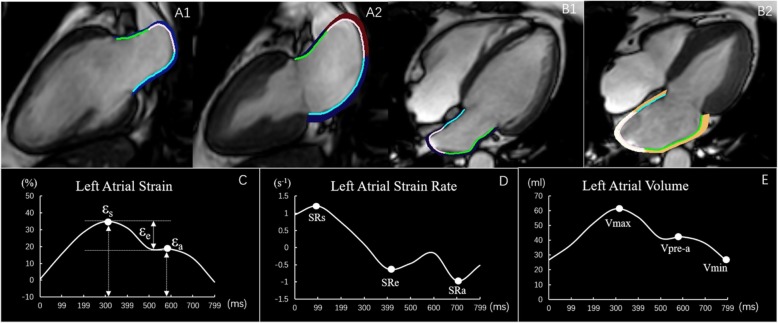
Fig. 1.
This figure shows a representative example of left atrial (LA) tracking on both the 2-and 4- chamber cines in a normal control subject. A1 and A2 left ventricular (LV) end-diastole and end-systole respectively on the 2-chamber view, B1and B2 LV end-diastole and end-systole respectively on the 4-chamber view. C and D The LA strain and strain rate curves. The total strain (εs), Passive strain (εe) and active strain (εa) were identified from the strain curves. The strain rates during LV systole (SRs), LV early diastole (SRe), and atrial contraction (SRa) were also determined from the strain rate curve. E LA volume curve. The LA maximum volume (Vmax), the pre-contraction volume (Vpre-a), and the minimum volume (Vmin) are shown here

