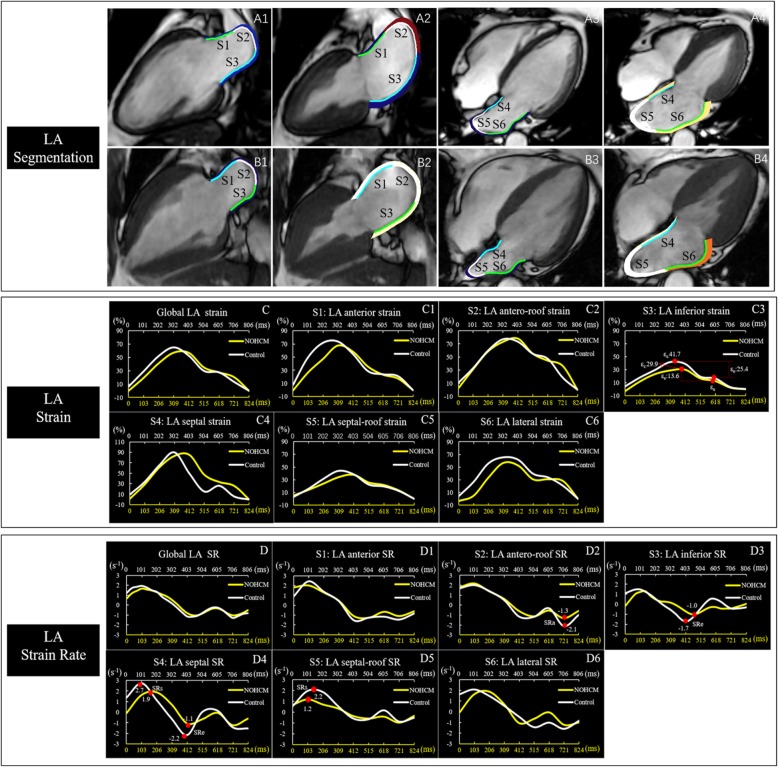
Fig. 2.
LA segmentation in representative cases of a healthy subject (A1–4) and a NOHCM patient (B1–4). The LA wall is automatically divided into 6 segments by the software [segment1(S1): anterior, segment2(S2): antero-roof, segment3(S3): inferior, segment4(S4): septal, segment5(S5): septal-roof, segment6(S6): lateral]. Comparison of LA global strain (C) and strain rate (D), segmental strain (C1–6) and strain rate (D1–6) between the non-obstructive hypertrophic cardiomyopathy (NOHCM) (yellow line) and the control (white line), the LA global strain and strain rate in the NOHCM were similar to the control, while segmental strain (inferior) and strain rate (antero-roof, inferior, septal and septal-roof) were lower in the NOHCM than the control. The yellow X axis represented the cardiac cycle length of a patient with NOHCM, and the white X axis represented the cardiac cycle length of a healthy control. εs = total strain, εe = passive strain, ε =, active strain, SRs = peak positive strain rat, SRe = peak early negative strain rate, SRa = peak late negative strain rate. Time dependent curves of the strain parameters were plotted offline using raw values provided by software

