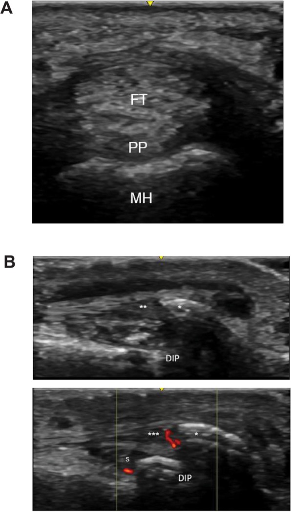
Fig. 3.

Ultrasound findings for differentiation of psoriatic arthritis from rheumatoid arthritis. a Short-axis view of palmar plate inflammation. FT, flexor tendon; MH, metacarpal head; PP, palmar plate. b Dorsal long view of enthesitis of the extensor tendon from a distal interphalangeal joint in a patient with psoriatic arthritis. DIP, distal interphalangeal; S, DIP synovitis; asterisk (*), enthesophyte; double asterisks (**), extensor tendon demonstrating thickening, hypoechogenicity, and loss of fibrillar architecture; triple asterisks (***), extensor tendon with insertional Doppler
