Fig. 5.
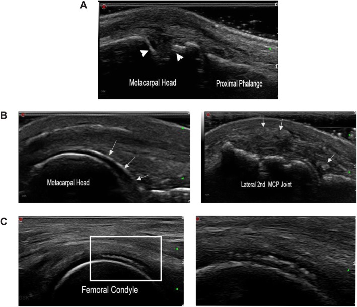
Ultrasound imaging of bone erosions and crystal deposits. a Transverse view of second metacarpophalangeal joint in a patient with rheumatoid arthritis; arrowheads denote bone erosion. b Left, chondrosynovial urate deposition at the second metacarpophalangeal joint (arrows); right, at the same joint, intra- and peri-articular tophaceous deposits seen as heterogeneous collections (arrows). c Left, calcium pyrophosphate crystal deposition seen sandwiched within the cartilage; right, magnified view of the white rectangular area on the left

